Rosacea treatment monitoring with skin ultrasound


Winter seems to have come back. Today I´ve been in Villalba, a mountain town,  in the speciality center with Monica, Derm nurse.  With her work seems also funnier and lighter… Thanks Monica.


In some ocasions, clinical evaluation of inflammatory conditions as rosacea is difficult only on clinical basis.

Ultrasonography allows evaluation of inflammatoy conditions in terms of B mode signs (dermal hypoechogenicity) and color Doppler (icrease or decrease of flow).

In the case I will show you our patient complained of nasal rosacea of torpid evolution.

We initiated doxicicline 100mg/bd  with much improvement

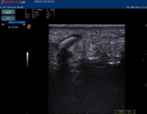
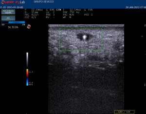
These two images correspond to initial and after treatment ultrasonography .

Clear decrease of edema and flow is evident.


_20120410203208_1628510

Pretreatment


rosacea post treatment

Post treatment


Plantar foreign bodies

This afternoon with  pleasent company and help of my colleague  Dr. Francsico  Javier Garcia Martinez (Last year Resident Dermatologist Santiago de Compostela  ) we have had an interesting case.

CLINICAL CASE

A 5 old year girl complained about plantar pain an nodule for  6 months.

The plantar nodule was evident and increased and decreased depending on weeks.

An ultrasound of the painful area was performed

_20130128203319_1715420_20130128203319_1718340

COMMENT OF CUTANEOUS US

  • A dermal-subdermal hyperchoic structure, surounded by a hypoechoic halo,  with posterior shadowing was evident.
  • Suspicion diagnosis of foreign body was stabished. Mother was explained about observation or extraction and conservative approach was adopted

TAKE HOME MESSAGE

  • Ultrasound is useful in the diagnosis of subcuraneous lesions
  • Artifacts as posterior acoustic shadowing are useful when establishing siagnosis of slid foreign bodies
  • Follow up and foreign body extraction is easier guided by ultrasound